.jpg)
.jpg)
.jpg)
.jpg)
Inscriete la noutatile noastre

Livrăm gratuit în raza municipiului Chișinău comenzile ce depășesc valoarea de 500 lei.
11 Nov 2020
Transplant integral de piele la o pisică cu avulsie cutanată podală distală - prezentare de caz
Full thickness mesh graft in a cat with degloving wound - case presentation
Florin Cristian Delureanu
First published: 20 noiembrie 2017
Editorial Group: MEDICHUB MEDIA
DOI: 10.26416/PV.29.8.2017.1252
Abstract
Applying skin grafts is one of the most common methods of closing distal defects in the limbs. At the same time, the results are also aesthetic. This paper shows the operatory steps present in applying a skin graft, approaching the wound before surgery to get granulation tissue and the postoperative care for a cat of about 5 years of age, with unknown history.
Keywords
skin mesh graft, degloving wound
Rezumat
Aplicarea grefelor de piele este una dintre cele mai frecvente metode de închidere a defectelor distale de la nivelul membrelor. În același timp, rezultatele obținute sunt și estetice. Această lucrare arată pașii operatori prezenți în aplicarea unei grefe de piele, abordarea plăgii înainte de intervenție pentru obținerea țesutului de granulație și îngrijirea postoperatorie la o pisică de aproximativ 5 ani, cu istoric necunoscut.
Cuvinte cheie
grefă de piele plagă prin avulsie
Introduction
An ample loss of skin with underlying tissue and exposure of deep components (e.g., tendons, ligaments, bones) define a degloving injury. These kinds of wounds are most frequently seen on distal limbs, medial tarsus/metatarsus. The main cause of degloving wounds is car accident, especially when the animal is dragged or pushed by a moving car. In all of the cases, bacteria and debris are present in the wound.
Free grafts are described as a piece of skin detached from an area of the body and placed over the wound. There are two types of free grafts when we talk about graft thickness:
full thickness (epidermis and entire dermis)
partial/split thickness (epidermis and a variable portion of dermis).
Skin grafts are used when there is a defect that cannot be closed by skin flaps or direct apposition. Two factors influence skin graft survival: revascularization and absorption of the tissue fluid.
Case report
A 4-year-old female, shorthair cat, weighting 3.25 kg, was presented to our clinic. Before that, the owner was at another clinic for consult and he was disappointed because they recommended euthanasia or amputation of the limb. Besides, the first veterinary doctor treated the cat with amoxi+clavulanate and Nekro Veyxym. The owner said that she went missing for about 10 days.
Clinical examination
After a thorough clinical exam, we found that all was normal, except for a degloving injury. The back right leg was affected.
There was a massive inflammation with infection and a lot of debris on the dorsal surface of metatarsal area (figure 1), and ventral above metatarsal pad.
.png)
Figure 1. Dorsal aspect of the metatarsal wound. Deep big tissue is affected; low to moderate discharge is present
Besides, also in the ventral metatarsal area, another wound located proximal to the metatarsal pad and 3 deep holes were identified at the base of second, third and fourth finger (figure 2). There could be distinguished the chronic aspect.
.png)
Figure 2. Ventral aspect of the wound. Note the swelling and the holes at the base of the fingers (red arrows)
Another lesion was noticed on the same leg, in the medial aspect of the thigh. This wound was deep with a circular shape (figure 3).
.png)
Figure 3. Deep wound with circular aspect, approximately 1.5 cm diameter, located near saphenous vein
We estimated that the lesion occurred about two weeks ago. We registered pain and high local temperature after palpation.
The cat was stable, normothermic, with normal color of mucous membrane, CRT 3 seconds and normal superficial lymph nodes.
Radiograph of the affected back limb
Two X-ray views were made to eliminate bone changes or foreign bodies (figure 4a, figure 4b).
.png)
Figure 4a, 4b. The specialist described: suspected slight thickening of phalanges cortical 1 fingers 3-4 and gently bending them. Soft tissue swelling of the tibio-tarso-metatarsian region
Approach
After evaluation, the initial recommendation included a good wound management under anesthesia. Before surgical debridement (figure 5a, 5b), culture was done.
.png)
Figure 5a, 5b. Dorsal and ventral aspect of the lesions after surgical debridment
Next, wound lavage was initiated with one bag of 500 ml of warm saline (the most easy way to deliver fluids on the wound is to connect the saline bag with a administration set to the syringe and needle with a 3-way stopcock; a large amount of liquid is needed to be effective).
Finally, this first stage ends with a wet-to-dry bandage. A primary wound closure was performed for the lesion placed on the medial aspect of the thigh (figure 6), after intensive cleaning, removal of foreign bodies and dead skin.
.png)
Figure 6. Wound closure by simple interrupted suture. 0-2 nonabsorbable material was used
Empirically, the cat received cefquinome until the results arrived and for pain management we administered tramadol 3 mg/kg and meloxicam 0.1-0.2 mg/kg. The cat recover well after anesthesia.
Culture result
One day before performing surgery, we received the culture result. Streptococcus canis (++++) was identified and was sensible to many antibiotics. Amoxicilin+clavulanic acid (Synulox® was initiated for general therapy and chloramphenicol ointment (Opticlor - Pasteur) for local therapy.
Next, a full thickness mesh graft was used on the dorsal aspect of the limb due to the length and depth of the wound, and the other wound was left for healing by second intention, both being protected by bandages. In the next 10 days, the limb wounds were treated in the same manner.
Removal of bacteria, granulation tissue formation and the beginning of epithelization were supported by next bandages as follows:
Day 1
Wet-to-dry bandage was used after surgical debridement (this kind of bandages adhere to the wound and remove the little layer of dead tissue when we take off). Soaked in warm saline 1-2 minutes before removing, they were changed after 24 hours one to the other. Cotton gauze was the primary contact layer of the bandage.
Day 2 and day 3
Moisture retentive dressing (MDR) - calcium alginate (Sorbalgon - Hartmann®) was the primary contact layer. It is good to use it when there exists high exudate like in our patient (figure 7a, 7b).
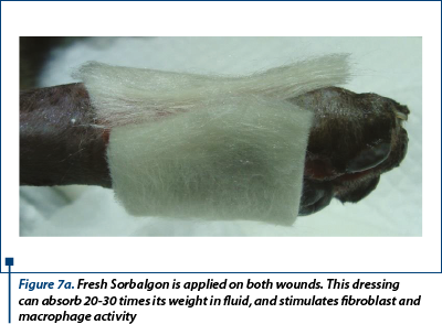
Figure 7a. Fresh Sorbalgon is applied on both wounds. This dressing can absorb 20-30 times its weight in fluid, and stimulates fibroblast and macrophage activity
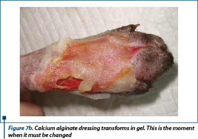
Figure 7b. Calcium alginate dressing transforms in gel. This is the moment when it must be changed
Day 4, 6 and day 9
Moisture retentive dressing (MDR) - hydrocolloid (Hydrocoll - Hartmann®) was the primary contact layer because the discharge decreased (figure 8).
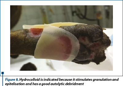
Figure 8. Hydrocolloid is indicated because it stimulates granulation and epitelisation and has a good autolytic debridment
After 9 days of management we inspected the wounds and we anticipated that in day 11 the granulation tissue will be ideal for performing the graft.
Day 11 - second anesthesia and surgery.
Describing surgical procedure
Preoperative surgical site preparation: the cat was placed in left lateral recumbency, with the wound exposed.
The limb was clipped entirely and povidone iodine and alcohol were used for aseptic surgery. Sterile warm saline 0.9% was used for wound lavage. Meanwhile, a colleague prepared the donor site - lower craniolateral thorax (right side) in the same manner.
Almost 1 mm of epithelium that has started to grow from the wound edges over the granulation tissue was removed using a thumb forceps and a no. 10 scalpel blade (figure 9).
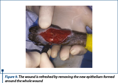
Figure 9. The wound is refreshed by removing the new epithelium formed around the whole wound
A perpendicular incision was made right at the edge of haired skin with epithelium. The wound was incised all around and after that the epithelium was removed by advancing the scalpel blade under the epithelium around the wound. Then, undermining was performed around the wound edges.
A fragment of sterile surgical drape was used over the wound to get the exact shape. The drape “pattern” was placed to the donor area.
To maintain the wound moist, we placed over a cotton gauze moistened in warm sterile saline 0.9% while the graft was transferred. The direction of hair growth was marked with a black arrow above the donor site, so that the direction of the hair growth on the graft would be the same as the hair growth direction on the skin surrounding the wound.
After that, the margins of the drape “pattern” was traced on the skin.
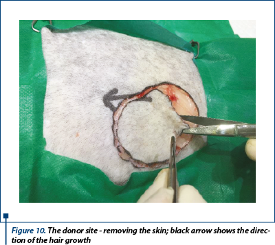
Figure 10. The donor site - removing the skin; black arrow shows the direction of the hair growth
The skin of the donor bed was incised with no. 10 scalpel blade and removed using thumb forceps and Metzenbaum scissors (figure 10). The defect left after removing the graft was primary closed by undermining and advancing the skin edges with walking sutures using 3-0 monofilament absorbable suture material and finally the skin was sutured in a simple interrupted suture using 2-0 monofilament nonabsorbable suture (figure 11a, 11b).
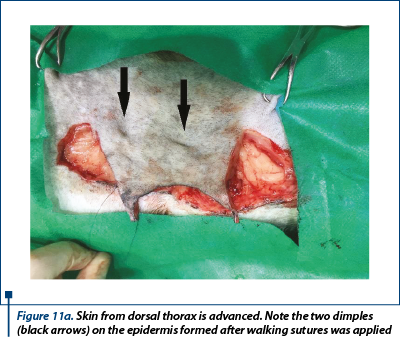
Figure 11a. Skin from dorsal thorax is advanced. Note the two dimples (black arrows) on the epidermis formed after walking sutures was applied
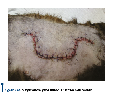
Figure 11b. Simple interrupted suture is used for skin closure
Preparing the graft
The dermal side of the graft was placed on a polystyrene board with a thickness of 10 cm covered with a sterile drape and after that we fixed and stretched with 21G needles. The subcutaneous tissue was removed from the graft. Next, parallel incisions was made in the graft, 0.5-0.7 cm long and apart (figure 12). At the end, the graft was placed on the granulation bed and sutured with 4-0 monofilament nonabsorbable suture in a simple interrupted suture manner. Additional tacking suture was placed to ensure the expansion of the mesh incision and allow the fluid drainage (figure 13).
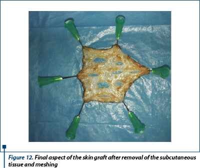
Figure 12. Final aspect of the skin graft after removal of the subcutaneous tissue and meshing
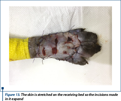
Figure 13. The skin is stretched on the receiving bed so the incisions made in it expand
Choosing the right bandage after grafting and aftercare
It is important to use a nonadherent primary dressing. My initial choice was Grassolind® (Hartmann). This dressing is sufficiently porous to allow easy passage of exudate from the wound surface and preventing maceration of surrounding tissue (figure 14). The ventral metatarsal wound maintain hydrocolloid dressing (Hydrocoll - Hartmann®) as primary layer. A thin layer of chloramphenicol oinment (Opticlor - Pasteur®) was used all around both wounds and over the graft.
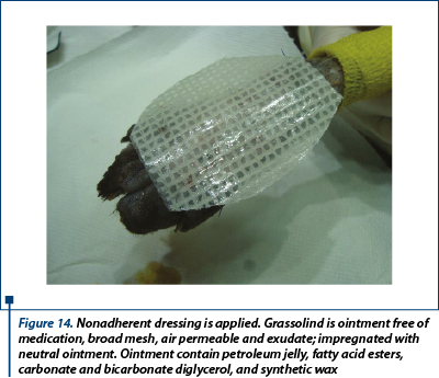
Figure 14. Nonadherent dressing is applied. Grassolind is ointment free of medication, broad mesh, air permeable and exudate; impregnated with neutral ointment. Ointment contain petroleum jelly, fatty acid esters, carbonate and bicarbonate diglycerol, and synthetic wax
Over the first dressings was applied 5 cm x 5 cm of compresses (Medicomp - Hartmann®) and a roll gauze was the second layer. After a few laps of gauze stirrups was placed to secure the bandage in place. Extemporaneous half “clamshell” splint (figure 15) was made from plastic material which was curved in such a way that the limb was fixed in semi flexion. The splint was a little bit longer than the extremity of the pelvic limb (“toe-dancing” position), thus provided a maximum relief pressure. In the proximal area, under the splint, I put cotton to prevent pressure injuries on the caudal aspect of the thigh. Applied from proximal to distal and with moderate tension, elastic warp was the final protective layer of the bandage and it was secured at the proximal end with tape.
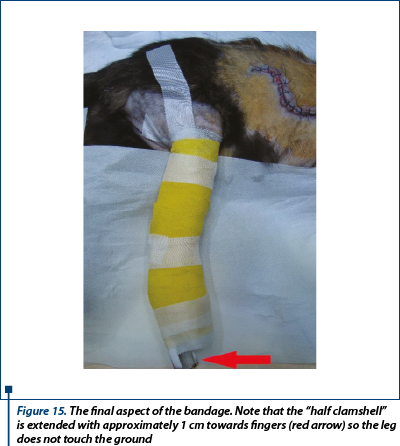
Figure 15. The final aspect of the bandage. Note that the “half clamshell” is extended with approximately 1 cm towards fingers (red arrow) so the leg does not touch the ground
Changing bandages
The bandage was changed in days 1, 3, 5, 7 and 10 postoperatively. In day 10 the suture material was removed from the graft and from the donor site. From day 17 to day 29 hydrogel (Hydrosorb - Hartmann®) was used as primary bandage layer and the bandage was changed from 4 to 4 days. In day 28 no discharge was present in the bandage; the wound was completely healed and 0.2-0.4 mm of hair was present in the center of the graft.

Figure 16. Macroscopic aspect of the ventral wound. Delayed healing on day 45 - epithelization stopped at this level
A delayed healing occurred at the wound in the ventral region (figure 16). From day 28 to day 59 epithelization has advanced very slow and granulation tissue has captured an appearance of ulcer (in this time, the wound was aseptic prepared and hydrocolloid and hydrogel were used as primary layer bandage and without the splint). In day 59 the wound was refreshed on the surface with a scalpel blade and laser therapy (figure 17a) and medical Manuka honey (figure 17b) was used daily for 14 days. After that, a complete healing was reached.
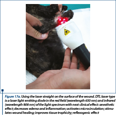
Figure 17a. Using the laser straight on the surface of the wound. DTL laser type is a laser light emitting diode in the red field (wavelength 650 nm) and infrared (wavelength 808 nm) of the light spectrum with next clinical effect: anesthetic effect; decreases edema and inflammation; activates microcirculation; stimulates wound healing; improves tissue trophicity; reflexogenic effect
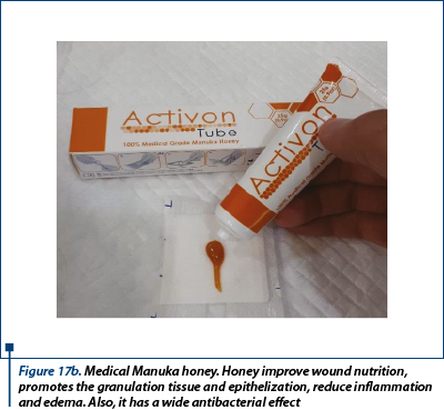
Figure 17b. Medical Manuka honey. Honey improve wound nutrition, promotes the granulation tissue and epithelization, reduce inflammation and edema. Also, it has a wide antibacterial effect
Illustrating wounds evolution after surgery
Day 1
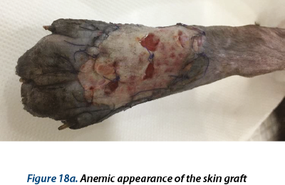
Figure 18a. Anemic appearance of the skin graft

Figure 18b. Normal granulation tissue and epitelization are present. The 3 holes located at the base of the fingers are healed
Day 11
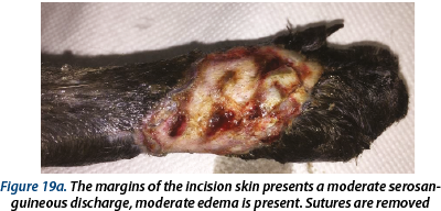
Figure 19a. The margins of the incision skin presents a moderate serosanguineous discharge, moderate edema is present. Sutures are removed
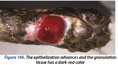
Figure 19b. The epithelization advances and the granulation tissue has a dark-red color
Day 28
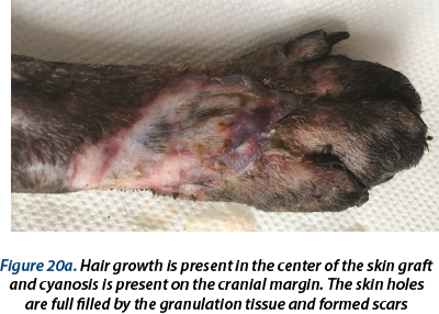
Figure 20a. Hair growth is present in the center of the skin graft and cyanosis is present on the cranial margin. The skin holes > are full filled by the granulation tissue and formed scars
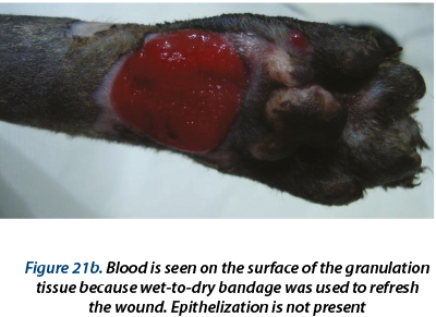
Figure 20b. The granulation tissue have a dry aspect
Day 35
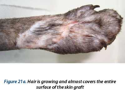
Figure 21a. Hair is growing and almost covers the entire surface of the skin graft
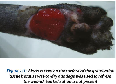
Figure 21b. Blood is seen on the surface of the granulation tissue because wet-to-dry bandage was used to refresh the wound. Epithelization is not present
Day 49
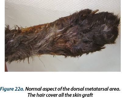
Figure 22a. Normal aspect of the dorsal metatarsal area. The hair cover all the skin graft
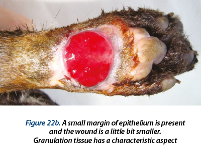
Figure 22b. A small margin of epithelium is present and the wound is a little bit smaller. Granulation tissue has a characteristic aspect
Day 11 after honey and laser therapyDay 16 after honey and laser therapy
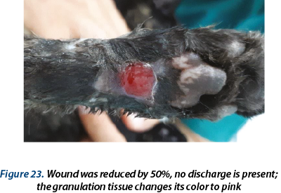
Figure 23. Wound was reduced by 50%, no discharge is present; the granulation tissue changes its color to pink
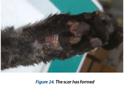
Figure 24. The scar has formed
3 months after surgery
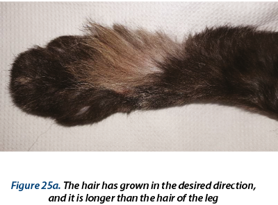
Figure 25a. The hair has grown in the desired direction, and it is longer than the hair of the leg
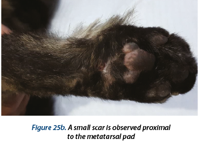
Figure 25b. A small scar is observed proximal to the metatarsal pad
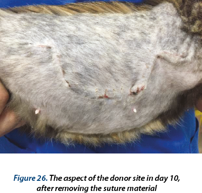
Figure 26. The aspect of the donor site in day 10, after removing the suture material
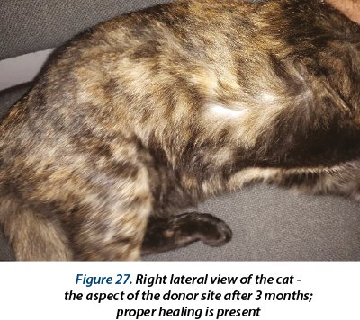
Figure 27. Right lateral view of the cat - the aspect of the donor site after 3 months; proper healing is present
Bibliografie
1. Steve F. Swaim, DVM, MS; Janet Welch, DVM, DACVS; Robert L. Gillette, DVM, MSE (2015) - Management of Small Animals Distal Limb Injuries, by Tenton NewMedia, USA.
2. Michael M. Pavletic, DVM - Atlas of Small Animal Wound Management and Reconstructive Surgery, Third Edition (2010), W.B. Saunders, USA.
3. Karen M. Tobias, DVM, MS, DACVS – Veterinary Surgery Small Animal (2012), Saunders , USA.
Sursă : https://www.medichub.ro/reviste-de-specialitate/practica-veterinara-ro/transplant-integral-de-piele-la-o-pisica-cu-avulsie-cutanata-podala-distala-prezentare-de-caz-id-1252-cmsid-69
Sep
05
Nefrolitiaza reprezintă o formă particulară de urolitiază, care poate evolua izolat sau asociată altor afecțiuni ale tractului urinar. Acest studiu a urmărit evaluarea aspectelor clinice, imagistice și terapeutice ale nefrolitiazei singulare la feline. Dintre cele 11 cazuri investigate, majoritatea pacienților au prezentat modificări clinice minime sau absente, diagnosticul fiind stabilit în principal prin examinare ultrasonografică. Opt cazuri (72,7%) au fost gestionate conservator, prin monitorizare clinică, imagistică și biochimică periodică, fără deteriorarea semnificativă a funcției renale. Trei pisici (27,3%) au necesitat nefrectomie, ca urmare a instalării sindromului de retenție azotată de gradul IV. Evoluția postoperatorie a fost favorabilă, cu ameliorarea parametrilor biochimici și stabilizarea funcției renale. Rezultatele obținute susțin faptul că majoritatea cazurilor de nefrolitiază singulară pot beneficia de management conservator, intervenția chirurgicală fiind rezervată cazurilor complicate, asociate cu afectare renală severă.
Aug
31
Ficatul reprezintă un organ vital pentru carnivorele domestice, având multiple funcții în cadrul organismului, prin intervenția în realizarea digestiei (secreția de bilă), înmagazinarea energiei sub formă de glicogen, filtrarea toxinelor de la nivelul circulației sanguine, sinteza de hormoni și proteine sau rol imunitar. Având în vedere capacitatea acestui organ de a se regenera, chiar și în condițiile unor leziuni extinse, semnele clinice manifestate de animale sunt deseori atipice sau apar în stadiile tardive ale evoluției diferitelor afecțiuni. Din aceste considerente, diagnosticul hepatopatiilor este adesea dificil de realizat și necesită o abordare integrată, care să includă date ale istoricului medical al pacientului, rezultatele examenului clinic, dar și o serie de investigații paraclinice – cum ar fi analizele de sânge (hematologie și biochimie), ecografie și radiografie abdominală, însoțite de puncție hepatică și examen citologic. Acest articol include patru cazuri ale unor pacienți (doi câini și două pisici) diagnosticați cu hepatopatii de etiologii diverse, în cadrul unui cabinet privat din București (Biji’s Animal Vet) în anul 2025, printr-un protocol de examinare care a inclus integrarea datelor rezultate din anamneză, examen clinic, analize de sânge, ecografie, radiografie și, pentru o parte din cazuri, examen citologic. Datele obținute au fost analizate și comparate cu cele furnizate de alte studii similare.
Jul
06
Ca entitate patologică asociată preponderent practicii medicale veterinare feline, gingivostomatita cronică reprezintă o afecțiune cu etiologie plurifactorială și tablou clinic dominat de prezența leziunilor de tip ulcerativ, dureroase și, adesea, cu dispunere bilaterală. Odată stabilit diagnosticul de certitudine, în cazul gingivostomatitei feline, urmărirea unei strategii țintite de planificare a protocolului terapeutic crește probabilitatea de a obține un rezultat pozitiv pe termen lung. Planul terapeutic este, astfel, menit să asigure o îmbunătățire netă a stării de sănătate, cu reale beneficii deopotrivă pentru pacientul felin și proprietar, și poate cuprinde, în primă fază, tratamentul medicamentos, ulterior recurgând la tratament chirurgical în cazurile refractare, de tip recidivant. În acest studiu ne propunem să evidențiem eficacitatea terapiei chirurgicale, ca ultim resort, în cazurile complexe de gingivostomatită felină, precum și o evidențiere a principalelor coordonate terapeutice de actualitate utilizate în managementul acestei patologii feline.
Jun
01
Gingivostomatita cronică felină reprezintă o afecțiune adesea observată în practica clinică veterinară. Caracterizată prin leziuni de tip ulcerativ, dispuse tipic bilateral, această patologie provoacă adesea durere și disconfort oral intens la pacienții felini afectați, consecințele clinice directe fiind disfagia și anorexia consecutivă, ce conduc la alterarea graduală a stării generale. Cu o etiologie multifactorială, incomplet elucidată, diagnosticul gingivostomatitei feline se bazează, în principal, pe tabloul clinic, completat de investigații paraclinice uzuale și specifice. Pacienții felini cu gingivostomatită cronică necesită studierea etiopatogeniei, scopul final fiind acela de a stabili, pe cât posibil, un diagnostic de certitudine. Un astfel de protocol de diagnostic include testarea pentru patologii precum FIV, FeLV și FCV, un set complet de analize hematologice și biochimice sangvine, precum și examene radiologice ale cavității bucale, fiind completat de biopsie și examinare microscopică, histopatologică, a țesuturilor afectate, care indică adesea prezența infiltratului limfoplasmocitar, sugerând un răspuns imunologic exacerbat. Astfel, posibile anomalii ale sistemului imunitar, precum prezența bolilor autoimune, imunodeficiența felină, neoplazii etc., modifică răspunsul imun al pacientului și duc la apariția infecțiilor oportuniste, care contribuie la natura cronică pe care adesea o îmbracă această patologie. Examinarea radiografică este, de asemenea, esențială pentru evaluarea structurilor dentare, evidențierea gradului de extindere a bolii parodontale, evaluarea gradului de resorbție radiculară, pentru a decela eventuale procese neoplazice, precum și pentru evaluarea pre- și postextracțională acolo unde este cazul. Lucrarea de față cuprinde o prezentare succintă a gingivostomatitei cronice, ca patologie felină de actualitate, cu accent asupra principalelor coordonate clinico-diagnostice de interes.
Inscriete la noutatile noastre